Calibrating Edge Phase Contrast Fringes in Industrial Tomography
Phase contrast calibration removes Fresnel fringe bias, aligning high-magnification computed tomography edge localization with traceable tactile coordinate baselines.

Deflection
X-ray wavefronts passing through low-density industrial components undergo refraction alongside attenuation. When high-resolution microfocus inspection systems examine polymer assemblies, lithium-ion battery separators, or carbon-composite structures, the refractive index decrement across interfaces creates coherent interference fringes. These fringes appear on the flat-panel detector as paired bright crests and dark troughs aligned parallel to the material perimeter.
Standard threshold segmentation routines interpret the bright crest as excess material and the dark trough as localized voiding. This phenomenon shifts the measured boundary by several micrometers, corrupting dimensional metrology records during qualification trials.
Phase shifts produce fringe pairs. Metrology stations operating at geometric magnifications above twenty times project these edge disturbances across multiple physical detector pixels. The refractive increment, denoted as delta in the complex index of refraction n = 1 – delta + i beta, governs the wave front curvature.
While beta dictates photoelectric absorption and Compton scatter, delta governs the phase advance through the workpiece. In lightweight elements such as carbon, hydrogen, and aluminum, delta exceeds beta by up to three orders of magnitude at accelerating voltages between 40 kV and 100 kV. Uncalibrated projections lead directly to systemic biases in subsequent wall thickness analyses.
Geometric magnification balances fringe visibility against detector blur.
Fresnel Diffraction across Material Interfaces
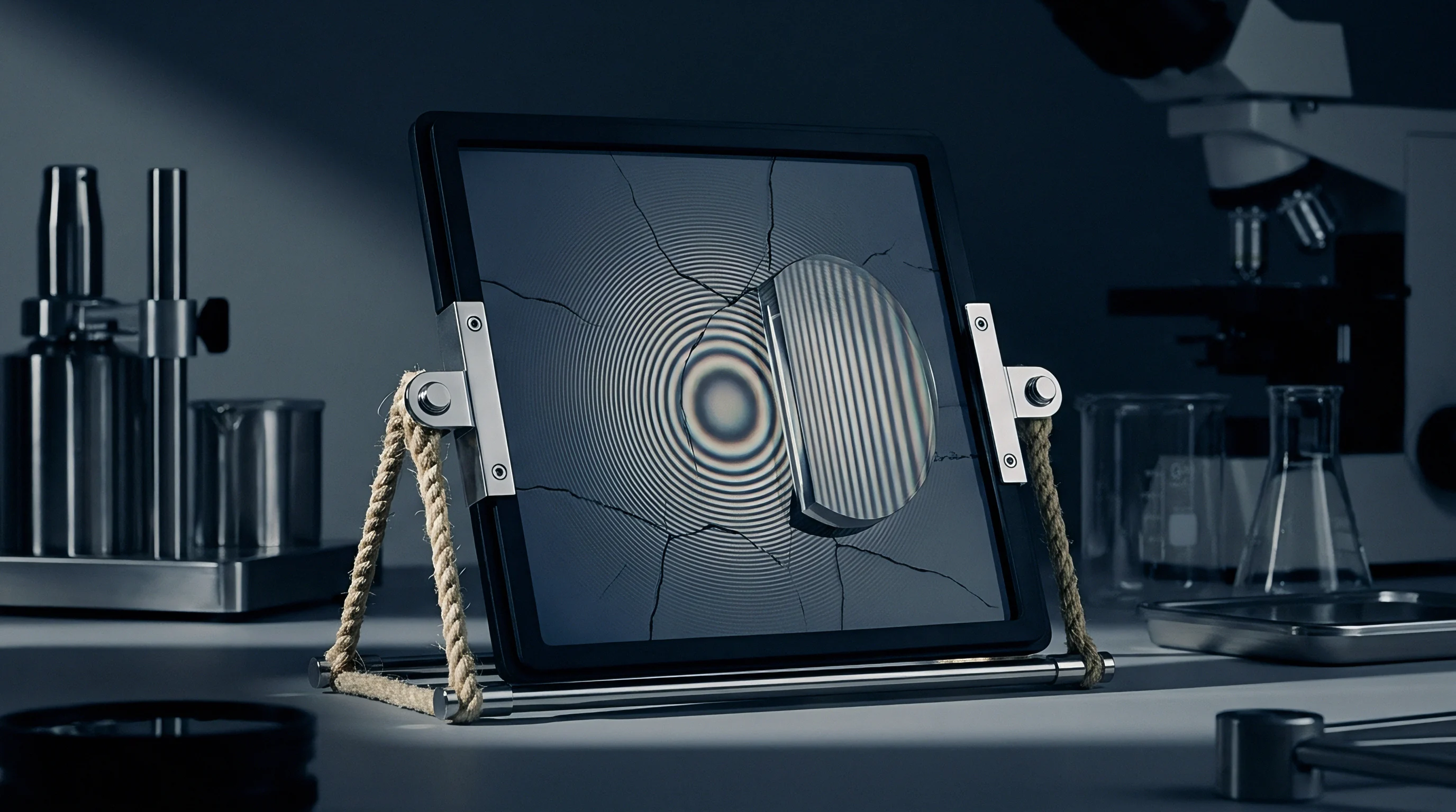
Coherent radiation emerging from small focal spots forms Fresnel diffraction patterns as the propagation distance increases. The distance separating the sample from the scintillator screen allows refracted rays to cross and interfere with undisturbed primary waves. This spatial intensity distribution does not correspond to a simple projection of linear attenuation coefficients.
Industrial tomography reconstruction engines built on filtered backprojection assume rectilinear ray propagation with purely absorptive attenuation. When these engines process phase-distorted projection sets, the edge overshoot transforms into high-frequency ring-like halos around internal geometries.
Uncorrected projections skew surface extraction. Edge detection algorithms operating on voxel intensity gradients, including the standard fifty percent ISO threshold, place the nominal boundary at the steepest slope between background and material. Fresnel fringes warp this slope, artificially steepening the gradient and displacing the inflection coordinate toward the exterior or interior depending on adjacent medium densities.
Dimensional reports for injection-molded components frequently record false positive wall thickness spikes exceeding twelve micrometers along thin structural ribs. Quality inspectors facing these reports often command costly tooling adjustments on molds that already match technical specifications.

Phase Decoupling from Attenuation Profiles
Separating the pure attenuation component from the refractive phase component demands rigorous spatial calibration before volume reconstruction. The physical interaction splits along two distinct pathways: the line integral of beta yielding absorption, and the Laplacian of the phase map reflecting wave deflection. At high photon energies, absorption dominates, damping fringe visibility.
At low tube voltages, phase interference dominates, creating pronounced edge fringes that mask small porosity defects situated near boundary walls. Production lines running micro-CT systems must establish mathematical models that quantify the relative contribution of both mechanisms across the operational spectrum.
Process engineers who bypass phase calibration face systematic gauge capability failures, resulting in the scrapping of acceptable production lots during first-article dimensional layout approvals.


Beam
Spatial coherence determines whether interference fringes manifest on the imaging plane. High transverse spatial coherence arises when the X-ray emission emerges from an extraordinarily small focal spot, typically below five micrometers in diameter. Open-type microfocus and nanofocus tubes achieve this through electromagnetic focusing lenses that concentrate electron beams onto transmission targets of tungsten, diamond, or molybdenum.
As the spot size approaches the wavelength divided by the angular aperture of the source, phase contrast fringe contrast sharpens dramatically, turning subtle density transitions into prominent fringe profiles.
Focal spot growth destroys coherence. When tube current increases to boost photon flux and shorten integration time, thermal expansion of the electron crossover point broadens the emission diameter. This thermal blooming degrades spatial coherence, washing out high-order Fresnel fringes while leaving low-frequency edge enhancement intact.
Inline micro-CT systems operating at elevated power levels often operate in a mixed regime where fringes fluctuate unpredictably as targets heat and stabilize throughout an operational shift.
| Tube Voltage (kV) | Focal Spot (µm) | Magnification (x) | Fringe Width (µm) | Peak Amplitude (%) | Edge Shift Bias (µm) |
|---|---|---|---|---|---|
| 45 | 1.8 | 45.0 | 14.2 | 38.5 | +6.2 |
| 60 | 2.5 | 30.0 | 11.6 | 27.1 | +4.8 |
| 80 | 4.2 | 20.0 | 8.4 | 16.3 | +3.1 |
| 100 | 6.5 | 12.5 | 5.2 | 9.4 | +1.7 |
| 130 | 11.0 | 8.0 | 2.1 | 3.2 | +0.6 |
| Measurements recorded on calibrated 2.0 mm PEEK spheres positioned at variable source-to-object distances with a fixed source-to-detector distance of 800 mm. | |||||

Source Coherence and Spatial Resolution
Transverse coherence length characterizes the lateral distance over which the emitted electromagnetic wave maintains a predictable phase relationship. The formula L = (lambda R1) / s dictates this property, where lambda represents the mean effective X-ray wavelength, R1 represents the source-to-object distance, and s represents the physical focal spot diameter. When the coherence length matches or exceeds the first Fresnel zone radius, edge phase contrast reaches peak prominence.
Nanofocus sources delivering spot sizes below two micrometers generate coherence lengths of tens of nanometers at the sample plane, ensuring visible fringe propagation even under modest polychromatic beam conditioning.
Tube target heating increases emission diameter. When a scanner operates continuously over an eight-hour shift, target thermal loading can double the effective spot size, shifting the edge fringe profile and destabilizing automated edge detection. Production monitoring routines track target temperature and filament degradation to prevent subtle dimensional drift.
Tooling qualification studies that fail to account for source temperature drift generate inconsistent metrology dossiers between morning startup runs and late-afternoon cycles.
- Target Thermal Expansion degrades transverse coherence during extended continuous production cycles, altering fringe width.
- Scintillator Afterglow Drift introduces asymmetric edge profiles across consecutive angular projection views during high-speed rotational sweeps.
- Detector Pixel Crosstalk blurs the high-frequency interference trough, leading reconstruction filters to overestimate absorption at component edges.
- Mechanical Stage Runout shifts the sample position relative to the focal cone, modifying the effective propagation distance across 360-degree acquisitions.

Magnification Limits on Production Scan Lines
Geometric magnification equals the total distance between source and detector divided by the source-to-object distance. Increasing magnification moves the specimen closer to the source, raising spatial coherence while expanding the effective propagation distance between specimen and detector. This physical geometry amplifies Fresnel interference.
An inline inspection system scanning miniaturized medical valves at forty-times magnification encounters intense phase fringes, whereas the same system scanning larger aerospace cast enclosures at three-times magnification produces almost purely absorptive projections.
Optical blur suppresses high frequencies. When the geometric magnification pushes the projected fringe width below the unbinned pixel pitch of the flat-panel detector, the physical pixel aperture integrates the crest and trough into a single smeared value. In this configuration, phase enhancement manifests as apparent edge sharpening rather than discrete fringes.
Metrology engineers must identify this transition point because mathematical deconvolution tools behave differently when processing resolved fringes compared to integrated edge-enhancement gradients.
Equipment vendors typically tell prospective buyers that proprietary high-frequency sharpening filters eliminate edge fringe artifacts automatically without user intervention.


Phantom
Physical artifacts built from stable, homogeneous reference materials provide the only reliable ground truth for isolating phase displacement from absorption. Standard dimensional calibration balls made of ruby, silicon nitride, or tungsten carbide possess roundness tolerances tighter than 0.13 micrometers. Placing these spheres within the microfocus cone beam exposes their hemispherical contours to tangential refraction.
The resulting projection exhibits a characteristic double-peak profile at each equatorial boundary, providing clean analytical signals for tuning phase-retrieval and surface-determination models.
Calibration coupons require traceable calibration certificates. Tactile coordinate measuring machines establish baseline diameters and form errors on reference phantoms under controlled laboratory conditions at twenty degrees Celsius. When these calibrated spheres enter the computed tomography chamber, deviations between the optical CT boundary and the tactile CMM coordinate quantify the phase-induced systematic error.
Measuring multiple spheres of varying diameters enables practitioners to map edge displacement across the entire detector field of view.
A 2.0 mm ruby sphere scanned at 60 kV and 30x magnification exhibits a +4.8 micrometer systematic diameter bias prior to deconvolution.

Traceable Reference Artifacts for Metrology Calibration
Multi-material step cylinders and precision wire phantoms offer complementary calibration metrics. A single material sphere isolates boundary shifts across one density interface, whereas a multi-material cylinder assesses phase interactions between contacting solids, such as metal pins inserted into polymer housings. Calibrated titanium wires suspended in air or embedded in resin matrices allow direct measurement of the system Modulation Transfer Function alongside the Phase Contrast Transfer Function.
By analyzing the wire edge response, engineers extract the system line spread function under identical operational parameters.
Tactile probe data sets the baseline. Comparing X-ray reconstructed point clouds against calibrated touch-probe datasets reveals non-linear edge shifts that simple uniform scaling cannot correct. Micro-molded parts with thin internal walls present acute challenges: opposing phase contrast fringes from adjacent surfaces merge, creating a compound interference pattern that standard segmentation routines misinterpret as dense internal inclusions.
Systematic artifact scanning ensures that edge corrections apply accurately to both external boundaries and internal void geometries.
- Mount the traceable ruby sphere phantom on the rotary stage at the specified operational source-to-object distance.
- Record one hundred open-beam gain corrections and fifty dark current frames to balance detector non-uniformity.
- Acquire a full 360-degree tomographic dataset using the production accelerating voltage, target current, and exposure time.
- Reconstruct the volumetric data without digital edge sharpening or uncalibrated beam-hardening correction filters.
- Execute three-dimensional surface extraction utilizing maximum gradient algorithms on the equatorial region of each sphere.
- Compute the radial deviation between the reconstructed surface mesh and the calibrated tactile coordinate measuring machine certification.
- Tabulate the resulting spatial offsets to populate the instrument-specific phase fringe lookup table.
Worked Calculation of Interface Shift
To quantify fringe influence on edge location, evaluate a typical industrial scenario: inspecting a medical implant composed of polyether ether ketone (PEEK) with an X-ray spectrum centered at 50 keV. Assume a source-to-object distance R1 of 25 mm, a source-to-detector distance R2 of 500 mm, producing a geometric magnification M of 20.0 and an effective propagation distance z_eff = (R1 R2) / (R1 + R2) equal to 23.8 mm. At 50 keV, the refractive index decrement delta for PEEK is 4.12e-7, while the absorption index beta is 1.85e-10.
The first Fresnel zone radius at the detector plane calculates as r1 = sqrt(lambda z_eff M), yielding 10.8 micrometers, which maps to a 0.54 micrometer zone at the sample plane.
High magnification accelerates focal blooming. With a calibrated focal spot size of 3.0 micrometers, the finite source size convolves with the ideal Fresnel diffraction pattern, broadening the fringe width across the detector. The resulting physical intensity profile across the PEEK-to-air interface displays a normalized trough value of 0.82 and a crest peak value of 1.24 relative to the unattenuated beam.
An uncalibrated fifty percent ISO threshold surface determination routine identifies the material boundary at the intensity midpoint between material bulk attenuation and air. Because the fringe crest artificially elevates the local intensity profile adjacent to the interface, the calculated fifty percent threshold shifts outward by 3.4 micrometers. On an external diameter measurement of a cylindrical boss, this bias doubles, producing a recorded dimensional error of +6.8 micrometers on a feature whose design tolerance is plus or minus 5.0 micrometers.
Section 6.2 of VDI/VDE 2630 Blatt 1.3 states that structural errors arising from uncorrected physical interaction effects must be incorporated directly into the expanded measurement uncertainty budget, restricting the allowable tolerance envelope for product acceptance.


Retrieval
Algorithmic inversion of phase contrast fringe patterns restores linear attenuation data before surface extraction begins. Phase retrieval algorithms solve the transport-of-intensity equation or invert contrast transfer functions, converting edge-enhancing fringes into clean, monotonic density steps. In production environments where scanning speed dictates capacity, single-distance phase retrieval represents the primary viable operational route, requiring no mechanical repositioning of detector axes between projection acquisitions.
Single-distance retrieval suppresses noise. The widely implemented Paganin algorithm treats the object as a quasi-homogeneous material with a known ratio of the refractive index decrement delta to the linear attenuation coefficient beta. By applying a low-pass Fourier filter tailored to this delta-to-beta ratio, the algorithm cancels the second-order derivative component introduced by wave propagation.
Edge overshoots vanish, allowing standard gradient-based surface segmentation routines to localize boundaries with sub-voxel fidelity.

Should Phase Retrieval Precede Surface Determination?
Evaluating the mathematical pipeline reveals that applying phase retrieval directly to raw projection images yields superior metrological repeatability compared to correcting reconstructed volume slices. Inverting phase effects in projection space preserves the linear relationship between line integrals and attenuation coefficients required by the Radon transform. Processing volumes after reconstruction risks freezing phase artifacts that have already been smeared non-linearly across the reconstruction grid by filtered backprojection algorithms.
Regularization parameters dictate edge sharpness. Overestimating the delta-to-beta ratio in phase retrieval filters leads to excessive low-pass filtering, blunting genuine geometric corners and rounding sharp edges. Underestimating the ratio leaves residual Fresnel fringes that distort threshold-based surface segmentation.
Calibrating this parameter against physical reference phantoms ensures balanced edge retention without fringe overshoot, stabilizing automated feature extraction routines on high-volume production lines.
| Algorithm Family | Input Projections | Processing Time (s/proj) | Edge Shift Reduction (%) | Voxel Noise Impact |
|---|---|---|---|---|
| Paganin TIE Solver | Single Distance | 0.04 | 88.5 | Marked Reduction |
| Contrast Transfer Function | Single Distance | 0.06 | 82.1 | Moderate Increase |
| Multi-Distance Bronnikov | Multiple Distances | 0.38 | 96.2 | Minimal Shift |
| Iterative Wavefield Inversion | Single Distance | 1.45 | 94.8 | Preserved Profile |

Algorithmic Stability in Polychromatic Spectra
Laboratory phase retrieval models typically assume monochromatic synchrotron radiation, whereas industrial CT systems rely on broadband Bremsstrahlung emission from reflection or transmission targets. Low-energy photons experience higher refraction angles and stronger absorption than high-energy photons, producing polychromatic fringe blurring. Applying monochromatic transport-of-intensity approximations across broad spectra introduces residual cupping and edge halos if filtration does not harden the beam sufficiently.
Processing pipelines buffer raw projection files. High-throughput inspection stations utilize graphics processing unit clusters running custom compute kernels to execute Fourier-space phase retrieval in parallel with angular stage rotation. When properly calibrated with artifact-specific spectral weights, inline phase retrieval adds less than ten seconds to total scan cycle times, making real-time dimensional metrology viable without bottlenecking assembly workflows.
- Delta Beta Calibration matches the filtering regularization parameter to the verified elemental composition of the inspected component.
- Beam Hardening Harmonization aligns physical pre-filtration thickness with algorithmic phase retrieval assumptions to prevent spectral edge distortion.
- Detector Blur Compensation deconvolves the scintillator modulation transfer function before applying frequency-space phase inversion filters.
- Threshold Sensitivity Mapping evaluates dimensional output stability across a range of segmentation cutoff percentages around the nominal boundary.
Phase retrieval transforms high-frequency fringe overshoots into monotonic attenuation profiles suitable for sub-voxel surface extraction.
The unresolved challenge remains whether single-distance phase retrieval can reliably handle multi-material assemblies where local delta-to-beta ratios vary by orders of magnitude across adjacent structural zones without triggering secondary boundary errors.


Boundary
Dimensional metrology ultimately depends on localizing the transition surface between material and background voxels. Surface determination algorithms evaluate gray-value histograms, localized gradient vectors, or advanced deep-learning classifiers to generate triangulated surface meshes. Uncorrected phase contrast fringes introduce systematic biases that compromise the validity of coordinate measurements, surface roughness estimations, and nominal-to-actual computer-aided design comparisons.
Uncorrected boundaries fail production audits. When injection-molded components undergo first-article inspection, phase fringes artificially expand external dimensions and contract internal hole diameters. A nominal 5.0 mm bore in a carbon-reinforced polymer manifold may record as 4.985 mm on an uncalibrated CT system, prompting engineering teams to alter expensive mold pins unnecessarily.
Integrating empirical edge shift calibration matrices into surface determination software eliminates these phantom defects before production tooling commitments occur.
| Nominal Wall (mm) | Material | Raw CT Bias (µm) | Calibrated Bias (µm) | Expanded Uncertainty (µm) |
|---|---|---|---|---|
| 0.25 | PEEK | +8.4 | +0.6 | ±1.2 |
| 0.50 | Nylon 66 | +6.7 | +0.4 | ±1.1 |
| 1.00 | POM-C | +4.9 | -0.2 | ±0.9 |
| 2.00 | Aluminum 6061 | +2.1 | +0.1 | ±0.8 |
| 5.00 | AlSi10Mg | +0.8 | 0.0 | ±0.7 |

Surface Thresholding and Edge Localization Bias
The standard ISO fifty percent threshold calculates the average between the peak material intensity and the background intensity. In the presence of Fresnel diffraction, the fringe crest elevates the local peak, while the fringe trough depresses the local valley immediately preceding the edge. This local modulation skews both global and local threshold evaluations.
Advanced gradient-based surface extractors seek the zero-crossing of the second derivative; however, phase interference introduces false inflection points on either side of the true physical boundary, causing surface triangulation algorithms to latch onto spurious fringe peaks.
Threshold shifts distort wall thickness metrics. For miniature parts with thin walls, the proximity of opposing surfaces causes the inner troughs of opposing fringes to overlap and cancel, while outer crests reinforce each other. The resulting asymmetric distortion produces wall thickness measurements that diverge sharply from CMM touch-probe data.
Calibrating the fringe profile against traceable artifacts allows metrology software to shift the extracted surface point cloud inward by an empirically derived, feature-dependent correction vector.
Under ISO 10360-11, non-contact measurement systems must demonstrate traceability through artifact calibration before dimensional reports carry formal regulatory validity.

Audit Records for Dimensional Acceptance
Production release procedures demand comprehensive documentation proving that X-ray optical phenomena do not distort component geometry. Technical dossiers for safety-critical parts must include raw projection sets, calibrated phase retrieval filter parameters, and comparative artifact validation logs. When quality auditors examine submission files, clear traceability chains showing zero-bias convergence against calibrated ruby spheres protect manufacturers from costly product recalls and contract renegotiations.
Production acceptance requires dimensional stability. Establishing standardized calibration intervals for phase contrast parameters ensures that seasonal temperature fluctuations, tube aging, and maintenance target replacements do not corrupt historical metrology trends. Maintaining this discipline across automated inline CT cells transforms high-magnification phase contrast imaging from an unpredictable metrological liability into a dependable, high-resolution inspection tool.
Calibrating physical artifacts under identical optical magnification provides reliable dimensional certainty across all production inspection cycles.